

Unexpected discovery: from crystals to the cellular respiration of the malaria parasite
Researchers at the Bernhard Nocht Institute for Tropical Medicine (BNITM) have disproved a long-assumed function of a protein from the malaria parasite. For 20 years, it had been suspected that the heme detoxification protein (HDP) played a role in converting the iron-containing chemical compound heme into non-toxic crystals. However, the researchers have now revealed that this is not the case: HDP fulfills vital functions in the parasite’s respiratory chain and is not involved in the formation of hemozoin crystals. The findings were recently published in the journal Nature Communications.

The single-celled parasite Plasmodium causes malaria in humans. Mosquitoes of the genus Anopheles can transmit the parasite to humans through a bite. After a phase in the liver, the parasite invades the red blood cells of the human body to multiply. Inside these cells, the malaria pathogen builds a specialised structure known as a food vacuole. This structure acts like a digestive organ for the parasite, where it breaks down hemoglobin, the red pigment in human blood cells, to obtain essential nutrients. However, breaking down hemoglobin produces a problematic by-product: heme. This molecule becomes toxic to the parasite if it accumulates. To protect itself, the malaria pathogen converts the heme into an insoluble, crystalline form called hemozoin. This process is critical for the parasite’s survival.
“In my PhD thesis, I investigated the heme detoxification protein, or HDP for short. For over 20 years, in vitro experiments have shown that HDP plays a key role in converting toxic heme into harmless crystals. Until now, scientists assumed that the protein also carried out this function in the living malaria parasite. However, our latest findings show that the reality inside the parasite is different,” says Louis Sarrazin, a PhD student in the Molecular Parasitology BMFTR junior research group at BNITM.

The heme detoxification protein is not where it was expected to be
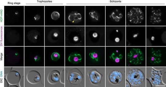
In an initial experiment, Louis Sarrazin aimed to determine the location of HDP within the malaria parasite. If it were involved in hemozoin crystal formation, it would need to be present in the food vacuole. To visualise the protein, he genetically modified it so that it fluoresces green. In living parasites, he could now see that the HDP was not found in the digestive vacuole, but instead in a different organelle: the mitochondrion. This was the first indication to the researchers that the protein fulfils a different function than previously thought.
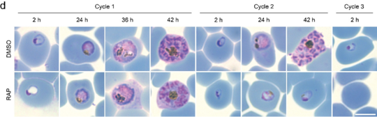
Next, Louis Sarrazin investigated whether hemozoin crystals would still form when he disrupted the HDP protein in the parasite. To do this, he genetically modified the parasites so that he could use a molecular switch to block HDP production. When no HDP was present in the parasite, it died and no hemozoin crystals were formed. But what was the cause and what was the effect? Did the crystals fail to form because the parasite died? Or was the crystal formation impaired, leading to the parasite’s death?
Why the parasite cannot survive without the heme detoxification protein
As the HDP was localised in the parasite’s mitochondria, the researchers focused on this organelle. Mitochondria are also found in human cells, where they perform the vital task of generating energy in the form of ATP through the so-called respiratory chain. In the malaria parasite, however, the mitochondrion does not function as a power station. During the parasite’s blood stage, the sole function of the respiratory chain is to remove electrons. These electrons would otherwise inhibit another pathway that is essential for the parasites’ reproduction. When the researchers inactivated the HDP in the parasite, the respiratory chain in the mitochondrion collapsed. As a result, the parasites were no longer able to replicate and ultimately died.

Dr Joachim Matz, head of the junior research group, explains the trick they used in the subsequent experiments to prevent the parasites from dying:
“We had to find a way to maintain the other pathway that is crucial for the parasite’s reproduction. Fortunately, yeast contains an enzyme capable of doing just that. We then introduced the genetic information for this yeast protein into the malaria parasites. This allows them to survive even when the respiratory chain in their mitochondria fails due to the absence of the HDP protein.”
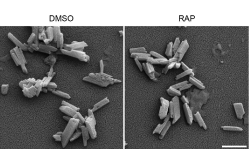
The strategy worked. Using this experimental approach, the team demonstrated that such “rescued” malaria parasites continued to form hemozoin crystals, even though the HDP protein was inactivated. This proves that HDP is not involved in crystal formation in living malaria parasites, as had been assumed. The essential function of HDP must therefore lie exclusively within the mitochondria.
How the heme detoxification protein keeps the respiratory chain running
To uncover the precise function of the HDP protein in the mitochondrion, Louis Sarrazin carried out computer modelling experiments. He discovered that HDP structurally resembles a specific protein from the Toxoplasma parasite. Toxoplasma and malaria parasites are closely related. The protein from the Toxoplasma parasite is a subunit of its mitochondrial ribosome. This ribosome translates the genetic information for components of the respiratory chain and synthesises the corresponding proteins. This led the researchers to suspect that HDP in the malaria parasite might play a role in mitochondrial protein synthesis. They therefore investigated whether inactivating HDP led to a reduction of certain proteins in the malaria parasite. This was indeed the case: proteins from respiratory chain subunits were present in much lower quantities than in the control malaria parasites. As a result, they can no longer fulfil their critical role of electron transport in the respiratory chain.
“Our study shows that even when a protein has been assigned a specific role for decades, it is worth taking a closer look. What appeared to be a crystal-forming agent in vitro turns out to be a vital component of the malaria parasite’s mitochondrial machinery in its living context. This discovery underlines how important it is to investigate fundamental processes within their native biological context. After all, only by understanding how the parasite functions can we identify its vulnerabilities and potential new targets for drug development,” says Louis Sarrazin, summarising his work.
Financial support was provided by, amongst others, the Federal Ministry of Research, Technology and Space (BMFTR), the German Research Foundation (DFG) and the Jürgen Manchot Foundation.
Original publication
Sarrazin L. et al., The Plasmodium heme detoxification protein functions in mitochondrial protein synthesis, Nature Communications 2026, DOI: https://doi.org/10.1038/s41467-026-76511-6
Contact person
Louis Sarrazin
Junior Group Molecular Parasitology
Phone : +49 40 285380-481
Email : louis.sarrazin@bnitm.de
Dr Joachim Michael Matz
Junior Group Leader Molecular Parasitology
Phone : +49 40 285380-301
Email : joachim.matz@bnitm.de
Dr Anna Hein
Public Relations
Phone : +49 40 285380-269
Email : presse@bnitm.de
Further information