Transformable down to the smallest cell structures
International research team succeeds in gaining insight into the molecular bag of tricks of the malaria pathogen Plasmodium falciparum
Malaria parasites have many faces: in the course of their life cycle, they develop several different cell forms. But how this metamorphosis takes place in detail is largely unclear. Molecular biologists from ten European research institutes have now used high-resolution imaging techniques to visualise the cell skeleton of several parasite forms. The surprising results have been published in the journal Nature Communications.

The malaria parasite is a true quick-change artist: In the course of its evolution, it has developed the ability to optimally use its two completely different hosts - humans and Anopheles mosquitoes - for its reproduction and spread. Depending on the tissue that the parasite passes through throughout its life cycle, it produces highly specialised and morphologically different cell forms that are optimally adapted to the respective host cells. How exactly these cell forms are constructed is, however, still largely unknown.
An international research team led by Prof. Dr Tim Gilberger, head of the Department of Cellular Parasitology at the Bernhard Nocht Institute for Tropical Medicine, and Prof. Dr Kay Grünewald, head of the Department of Structural Cell Biology of Viruses at the Leibniz Institute of Virology (LIV), has now analysed the molecular skeleton of four cell forms of the malaria parasite Plasmodium falciparum.
The researchers, whose laboratories are located in the CSSB (Centre for Structural Systems Biology) on the DESY campus in Bahrenfeld, were mainly interested in the structure of the microtubules: elongated structural elements made of proteins, so-called protofilaments. In all organisms with a cell nuclei, including humans, microtubules are made up of 13 protofilaments. This number was also assumed to be correct for the cell forms of unicellular parasites such as the malaria pathogen. Individual deviations were thought to be outlier phenomena.
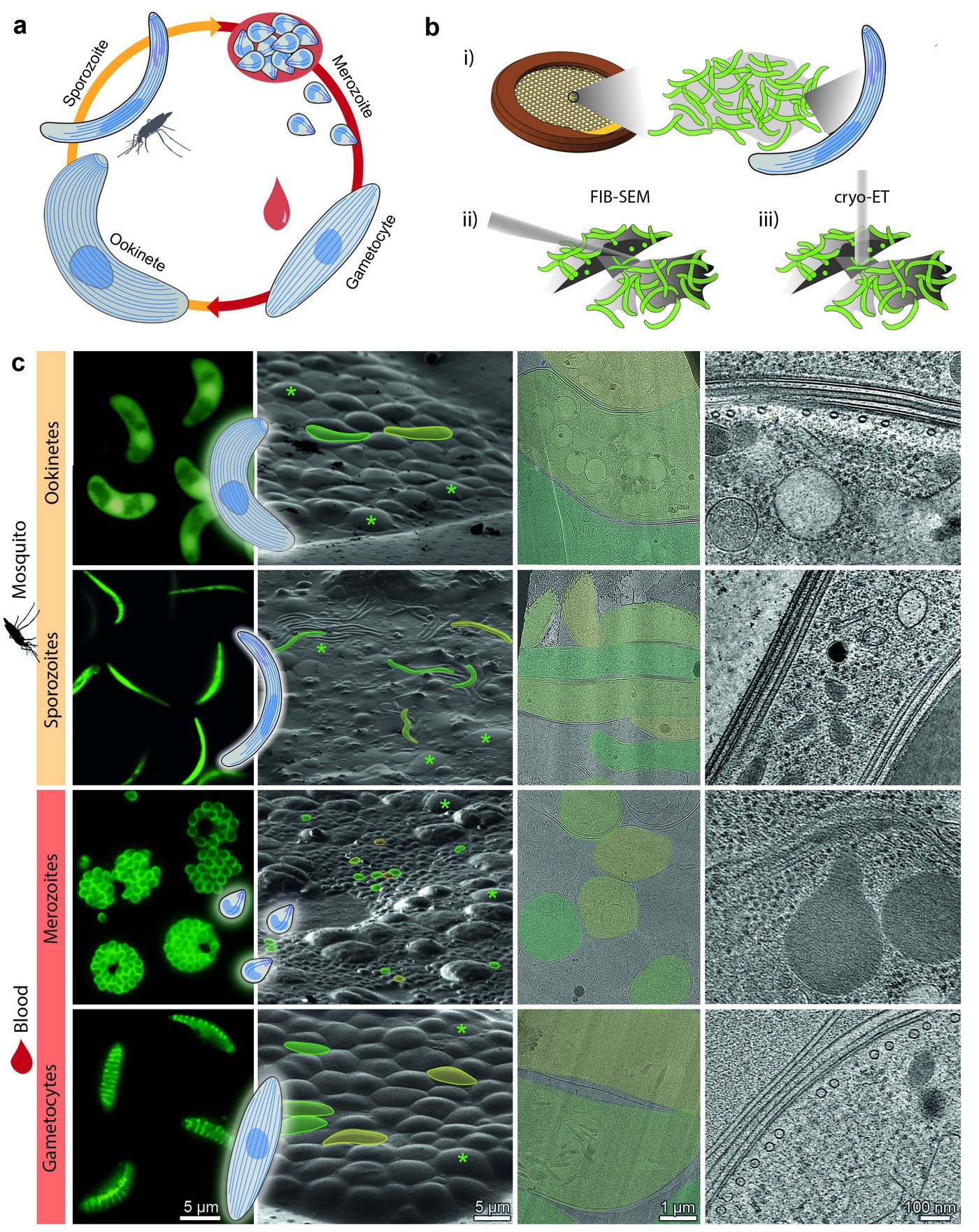
The research team decided to take a closer look at the microtubule architecture of the malaria pathogen and conducted comparative studies in four different parasite stages with the help of electron cryotomography. Could the key to the parasite's art of transformation lie in its microtubule?

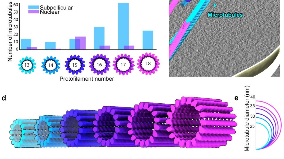
The data obtained revealed a spectacular diversity in microtubule architecture, not only from cell form to cell form, but also within a single cell form, namely in the gametocytes. These are the sexually differentiated malaria cells that the mosquito takes up from human blood during the mosquito bite. The banana-shaped gametocytes use microtubules composed of 13 to 18 protofilaments, which also form doublets, triplets and quadruplets. According to the study, such a large structural diversity is unprecedented and raises new questions regarding the underlying mechanisms and physiological significance.

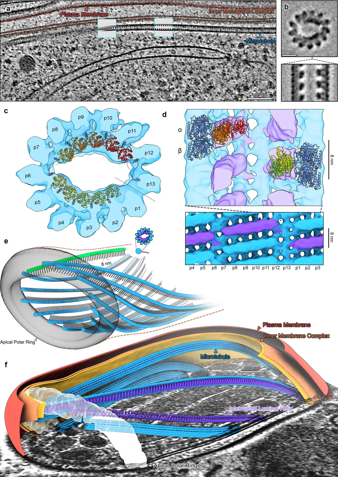
In merozoites, the malaria cell type that infects our red blood cells, the team observed the typical canonical microtubule shape with 13 protofilaments and was able to visualise other functional structures in detail due to the high resolution of the imaging technique used.
Sporozoites are the malaria cell forms in the saliva of the mosquito, which the mosquito injects into humans when it bites them and which then first migrate to their liver. Ookinetes result from the sexual reproduction of the malaria parasite in the mosquito's intestine: when the gametocytes have become gametes that fuse with each other.
The two parasite cell forms found in mosquitoes, sporozoites and ookinetes, also have the previously assumed microtubule structure with 13 protofilaments. However, in contrast to the merozoites, they again have a special feature: a filling of the cavity within the microtubules known as "interrupted luminal helices" (ILH). The research team suspects that the parasite's specially designed ILH provide stability without compromising flexibility. Balancing flexibility and stability is essential for the parasite's survival at all stages of malaria, and sometimes one is more in demand than the other: flexibility for the cell's dynamics or stability to keep the cell in shape.
"This diversity of microtubule structures has not yet been observed in any other organism in the species and will help to understand the parasite's art of transformation mechanistically," says Prof. Dr. Tim Gilberger.
The research team assumes that the actual diversity of microtubule structures of organisms with a cell nucleus is greater than currently assumed. With the imaging techniques now available, it is possible to represent and understand the dynamic architecture of many more cell types in detail.
Original publication
Tim-W. Gilberger & Kai Grünewald et al.: Variable microtubule architecture in the malaria parasite. Nature Communications 14, Article number: 1216 (2023).
Contact person
Prof. Dr Tim-Wolf Gilberger
Cellular Parasitology Department
Phone : +49 40 8998 87600
Email : gilberger@bnitm.de
Julia Rauner
Public Relations
Phone : +49 40 285380-264
Email : presse@bnitm.de


