Research Projects - Bacteriology
Epidemiology and immunology of arthropod-borne bacterial infections
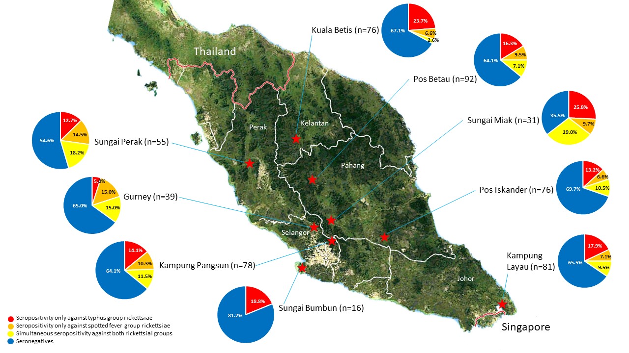
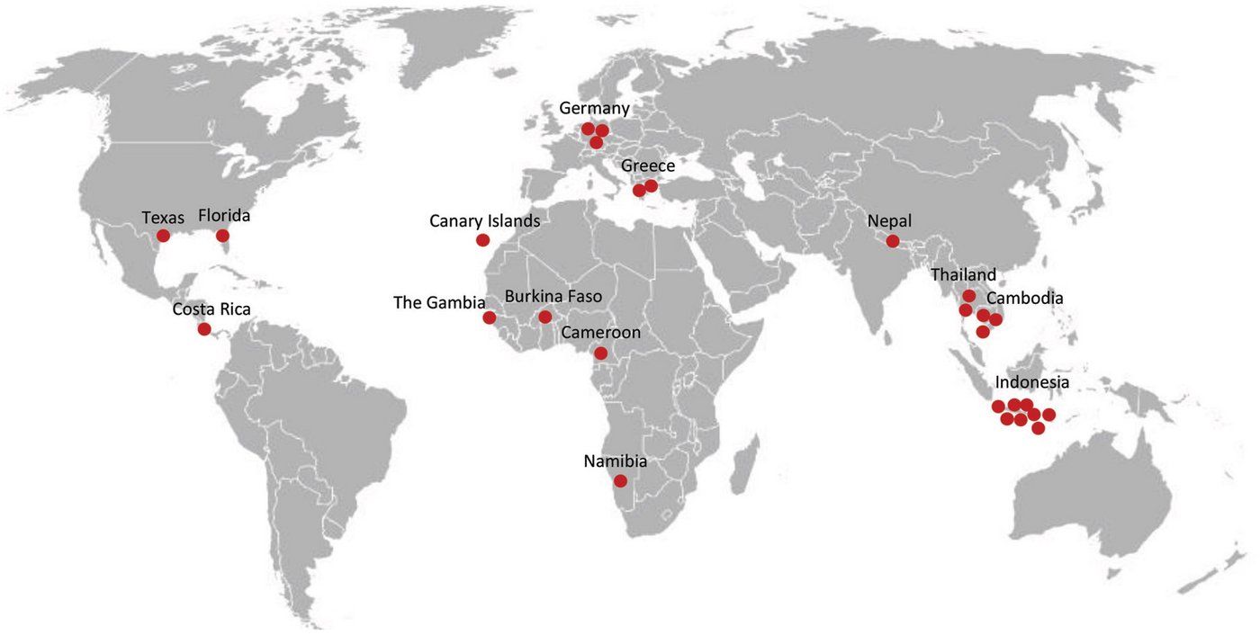
The research group investigates the epidemiology and immunology (antibody, cytokine and cellular responses) of arthropod-borne bacterial pathogens such as Rickettsia typhi and R. prowazekii, Orientia tsutsugamushi, Coxiella burnetii, Francisella tularensis, and Yersinia pestis. With a focus on rickettsioses, we conduct studies in Malaysia, Tanzania, the Democratic Republic of the Congo and Madagascar together with our international partners. Moreover, cohorts of returning travellers with various forms of rickettsioses, scrub typhus, as well as patients with autochthonously acquired tularemia and Q fever are analyzed.
For a list of the group's publications about rickettsioses, click here. More information (in German) about ticks and their respective pathogens which are diagnosed at our National Reference Center can be found here. For a list of the group's publications about ticks, see here. The research group's publications about scrub typhus can be found here, about tularemia here, and for contributions to Q fever research follow this link.
Epidemiological studies of rickettsioses
Typhus group rickettsiosis is caused by Rickettsia typhi and R. prowazekii. R. typhi, which causes murine typhus, the less severe endemic form of typhus, is transmitted by fleas; R. prowazekii, which causes the severe epidemic form of typhus, is transmitted by body lice. In contrast, the spotted fever group rickettsiae, such as R. africae and many other members of this group, are harbored by ticks.
Besides ongoing prevelance studies in the arthropod vectors, seroprevalence and cohort studies in humans are conducted. The occurrence of the pathogens in vectors and their prevalence in human disease are not always congruent and hint at local modifying factors.
For a list of our publications on rickettsial epidemiology, click here.
Immunological studies of rickettsioses
We analyzed patient cohorts with different forms of rickettsial disease caused by typhus group rickettsiae (R. prowazekii and R. typhi), spotted fever group rickettsiae (R. africae and others) and transitional group rickettsiae (R. felis), as well as the related scrub typhus caused by Orientia tsutsugamushi. Clinical data was evaluated and patient serum was examined for antibody kinetics, serum cytokine changes, and, in one cohort, also T cell responses.
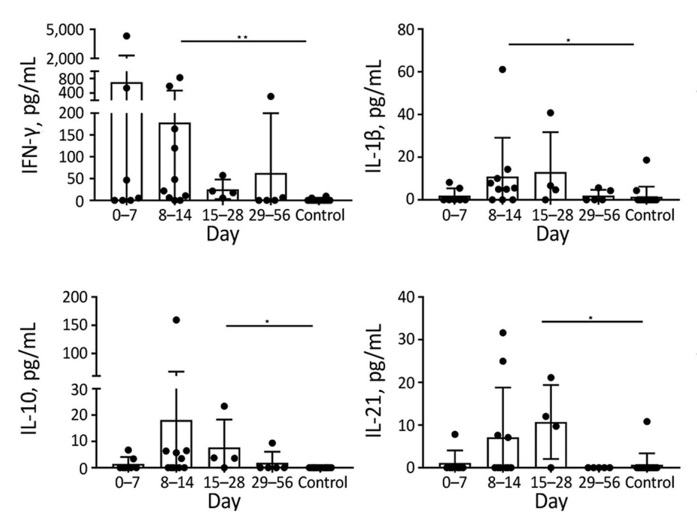
Strong cytokine responses were detected in the rickettsial typhus patient cohort. Cytokine levels peaked during the second and third week of infection, coinciding with seroconversion and organ dysfunction, respectively. In patients with the clinically similar scrub typhus, cytokine responses showed a rather mixed pattern.
For a list of the group's publications on rickettsial immunology, click here.
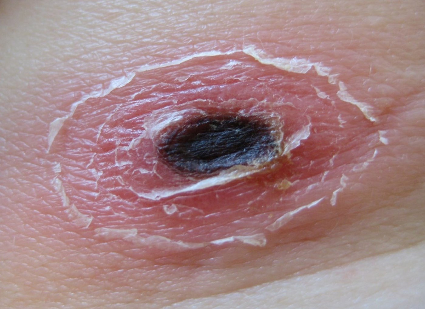
While eschars are often absent in R. typhi infections, the skin lesion can be found in African tick bite fever by R. africae (see above). These skin lesions can be used to detect the infecting pathogen molecularly and thus define patient cohorts specifically.
In typhus group rickettsiosis tissue reactions were examined (left), showing scattered nodules throughout organs involved. Besides the affected endothelial cells, the nodules consisted of activated CD4+ and CD8+ T cells and a surrounding microglial activation.
Further studies will investigate the cytokine response in tissues and brain organoid models using transcriptomics and individual cytokine mRNA measurements to elucidate pathogenesis and organ dysfunction.
Vector studies
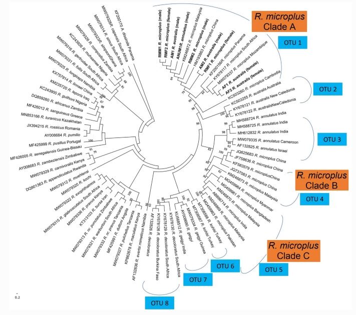
Together with our international partners, especially at the Universiti Teknologi MARA in Malaysia, we collect and identify tick species and analyze them for zoonotic bacterial pathogens. Farm animals are being screened nation-wide for the presence of various ticks which are collected and examined for rickettsiae, borreliae, and other bacteria.
For a list of our collaborative publications on ticks, click here.
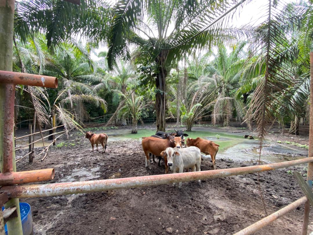
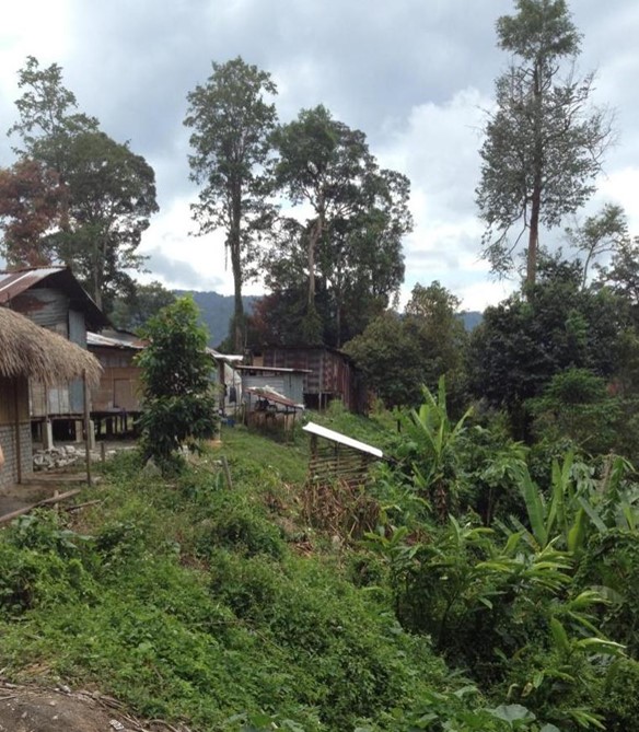
During the tick survey, multiple ectoparasite species were found on various hosts, and geographical mapping was performed. Tick species were characterized molecularly, and the spectrum of local tick species was expanded.


