


Alternativmethoden
Alternative methods
The scientists at the BNITM are keen to advance research into infection using methods that do not involve animal testing. In line with the 3R principle, these so-called alternative methods represent a combination of replacement and complementary approaches, which serve to reduce the number of animals used (reduce) and improve procedures involving animals (refine).
A key focus at the BNITM is the use of human organoids instead of conducting infection research on live animals. Organoids are three-dimensional clusters of cells grown in the laboratory that organise themselves into cellular structures, thereby resembling the development and organisation of tissues and organs in the human body. These in vitro miniaturised, simplified model systems of organs are derived either from reprogrammed pluripotent stem cells, which can form specialised cells such as liver cells through differentiation, or from healthy primary donor cells. These systems are generally based on findings from previous animal experiments.
At BNITM, we are currently using human organoids in several different studies on a wide range of parasitic and viral infectious diseases. For example, we use lung and/or liver organoids to identify mechanisms of the innate immune system that play a role in viral diseases such as influenza or parasitic diseases such as leishmaniasis. To gain better insights at the systemic biological level, initial infection models are also already being applied on multi-organ chips, on which different organoids are integrated into a physiological network. The application of these approaches may offer new potential strategies for treating these diseases in the future.
Although BNITM strives to reduce animal testing as a whole, it is not yet possible to dispense with animal testing entirely for certain research questions, as alternative methods have so far often only been able to simulate specific aspects of the complex processes occurring in the human body.
In order to further develop research in this field using innovative methods and technologies, the BNITM is part of the Leibniz Research Network ‘Stem Cells and Organoids’. Here, various institutes within the Leibniz Association pool their expertise so that these organ-like experimental models can serve as an additional alternative to animal testing.
Further information on the Leibniz Research Network can be found on the Leibniz Association’s website.
Amoebiasis
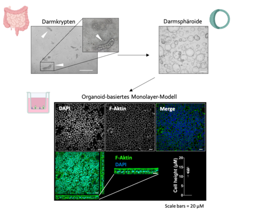
Spheroid-based monolayer model for investigating host-parasite interactions
Intestinal organoids are in vitro systems that mimic organ-specific structural and functional properties and can be generated from so-called spheroids. These ‘mini-intestines’ have a lumen and contain characteristic cell types of intestinal tissue. However, they are of limited suitability for infection studies involving the human pathogen Entamoeba histolytica, as the luminal side – the natural site of parasite invasion – is difficult to access. To nevertheless capitalise on the cellular complexity of the organoids, we use fragmented intestinal spheroids in our laboratory, which are cultured and differentiated as monolayers.
The monolayer model generated from intestinal spheroids is used to investigate the interaction between the parasite and the intestinal cells. In future, immune cells and a microbiome can be added to the system.
Funding: German Research Foundation (DFG)
Influenza
Organoid-based infection model for the analysis of inter-organ immune responses
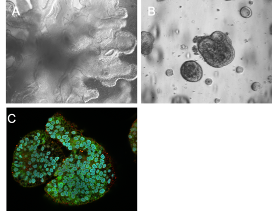
In this project, human pluripotent stem cells (iPS cells) are differentiated into functional lung and liver organoids using defined culture protocols that mimic key developmental signals through the use of stage-specific growth factors, extracellular matrix components and controlled environmental conditions. These organoids are incorporated into a so-called multi-organ chip system – a platform system in which the organoids are permanently connected to one another via microfluidic channels. This enables communication between the organoids via soluble mediators that are released in response to an infection.
The primary aim of this model is to infect the lung organoid with influenza viruses in order to trigger a local immune response and to investigate how this response affects the liver organoid via the chip’s shared circulatory system.
Funding: Leibniz Center Infection
Leishmaniasis
Organoids as host models for parasitic infections
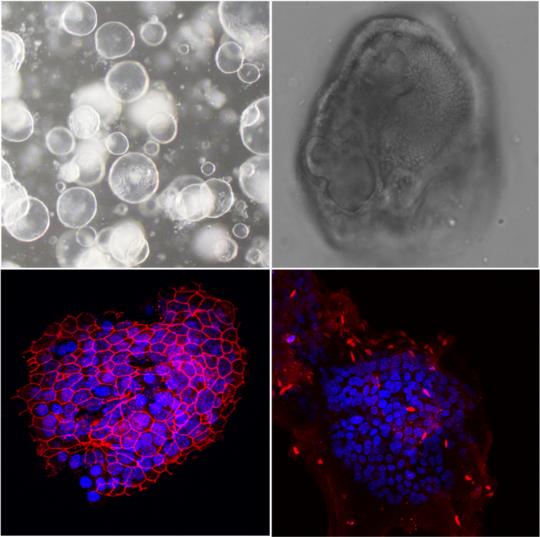
Parasitic infections of the liver are the subject of extensive research projects, but much of the knowledge gained is based on the use of mouse models. The advantages and disadvantages of these models have long been recognised. As an alternative, we use liver organoids in our research.
These are in vitro cultures that enable the long-term cultivation of human liver cells in a 3D structure. In this way, both the functional and structural properties of the liver can be replicated on a miniature scale. The liver cells required can be obtained from surplus tissue from biopsies or tumour resections.
We use liver organoids to model parasitic infections using human cells and to study their interaction with immune cells in the form of co-cultures.
Funding: Leibniz Center Infection
©BNITM | Melanie Lütkemeyer