


Exemplary animal models at the BNITM
Amoebiasis
Men are affected more frequently than women
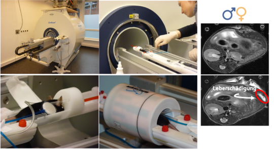
Women and men differ not only anatomically, but also in their immune responses. For example, men are more susceptible to various infectious diseases, whilst women are more prone to autoimmune diseases. However, the underlying biological processes are largely not understood. This is also evident in infections with the parasite Entamoeba histolytica, the causative agent of amoebiasis. The severe clinical form of the disease, known as amoebic liver abscess, mainly affects men. At the BNITM, a mouse model has been established that reflects these sex-specific differences. Following infection, male mice develop larger liver abscesses, whilst those in female mice heal more quickly.
Thanks to modern investigative methods such as magnetic resonance imaging, we are able to conduct experiments in accordance with the 3Rs principle as humanely as possible; furthermore, we have been able to significantly reduce the number of mice used in amoebiasis research.
Using this mouse model, we demonstrated that the male sex hormone testosterone is responsible for the gender difference via a direct influence on certain immune cells. Building on these findings, we were able to demonstrate that this mechanism of action is also applicable to human immune cells. Understanding these mechanisms paves the way for the development of new treatment strategies for infectious diseases that take the patient’s sex into account.
Further information can be found in the following publications:
Haemorrhagic Lassa fever
How does the immune response work?
Every year, there are approximately 300,000 new cases of Lassa fever in West Africa, caused by the highly pathogenic Lassa virus, of which around 5,000 are fatal. Making a correct diagnosis is challenging, as the initial symptoms of the disease – such as a sore throat, cough, chest and muscle pain, vomiting and diarrhoea – often cannot be distinguished from those of other illnesses such as typhoid or malaria. Furthermore, there are as yet no licensed vaccines or treatments for Lassa fever. This is partly because it is not yet known which factors within the human immune system play a role in the development of severe disease progression.
At BNITM, we have established a mouse model that can replicate the course of Lassa fever. It is used to examine various cell types of the immune system in more detail at different time points, in order to better understand their role in the disease process. We can also investigate whether immunomodulatory or antiviral drugs contribute to recovery.
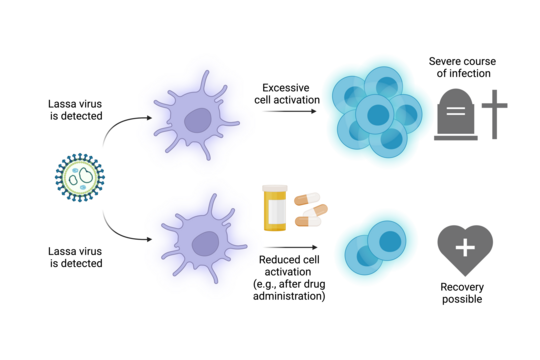
Using this mouse model, we have been able to demonstrate that the cells of the adaptive immune system are not adequately activated following a Lassa virus infection. Both an excessive immune response and one that is too slow and weak lead to a more severe course of the disease. Furthermore, we have shown that administering medication prevents the activation and excessive proliferation of immune cells, thereby enabling the host to survive the infection. Based on these findings, it is possible to develop therapeutic approaches for humans.
Further information can be found in the following publications:
Leishmaniasis
Research into immunomodulatory therapies
With an estimated 1.4 million new cases each year, leishmaniasis is one of the most significant parasitic infectious diseases worldwide, caused by protozoan pathogens of the genus Leishmania. Transmission occurs via the bite of the female sand fly. According to the World Health Organisation (WHO, 2018), leishmaniasis is endemic in 98 tropical and subtropical countries, with an estimated 350 million people acutely at risk from this poverty-related disease. However, due to globalisation, leishmaniasis is also becoming increasingly significant in other regions of the world, such as the Mediterranean and Central Europe.
Depending on the infecting Leishmania species and the host’s immune status, clinical symptoms range from cutaneous and mucocutaneous forms to fatal visceral leishmaniasis. Currently, no vaccines against leishmaniasis are available, and existing treatments have a number of drawbacks and risks. Cutaneous leishmaniasis is the most common form of the disease. It usually causes self-healing skin lesions that lead to disfiguring scars, often resulting in the stigmatisation of patients.
At BNITM, we use the mouse model of cutaneous leishmaniasis both to gain an immunological understanding of the parasite–host interaction and to establish new immunostimulatory treatment options. The mouse model replicates the clinical course of the disease as observed in patients and is therefore currently the most reliable choice for complex immunological studies as well as for studies on the efficacy of immunostimulatory drugs. We inject Leishmania parasites into the mice’s ears (simulating a sand fly bite), which after a few days leads to small to large, sometimes lesion-like swellings at the injection site; however, just as in humans, these heal spontaneously. The burden on the animals is considered to be low.
In leishmaniasis research at BNITM, pioneering methods are now emerging which offer promising and innovative alternatives to animal models for selected research questions.
Further information can be found in the following publication:
https://journals.asm.org/doi/10.1128/AAC.00161-20
You can find information on alternative methods under Alternative methods at BNITM.
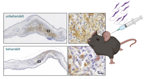
© Fehling et al. 2020 (created with biorender)