Tick bite and spotted fever
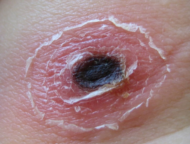
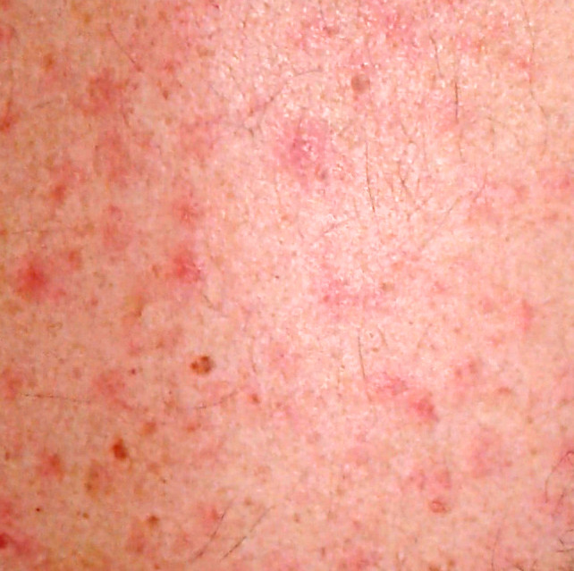
The BNITM offers various molecular biological and serological detection methods for the diagnosis of tick-bite fever and spotted fever (Examination orders bacterial disease, only in German). These diseases, triggered by various rickettsiae, often appear clinically as fever, headache, rash and a necrotic skin area, the eschar.
If these symptoms occur after a tick bite in the tropics, subtropics or also here in Germany, it is called tick-bite fever, which can be transmitted by different tick species.
Different tick-bite fever rickettsiae can trigger this disease. In Central Europe, for example, Rickettsia aeschlimannii, Rickettsia helvetica, Rickettsia slovaca, Rickettsia raoultii, Rickettsia monacensis are common. If infected in the Mediterranean region, Mediterranean tick-bite fever may be present due to Rickettsia conorii; if infected in South Africa, for example, African tick-bite fever may be present due to Rickettsia africae.

Tick-bite fevers are comparatively mild, with the exception of Rocky Mountain Spotted Fever, which is often severe and caused by Rickettsia rickettsii after a tick bite in North and South America or the Caribbean.

If the mentioned symptoms occur after a clothes louse infestation or flea bites, but often without eschar, its called epidemic or endemic spotted fever. The exanthema looks similar to the rash in tick-bite fever.
The spotted fevers are a much more serious diseases, in which complications such as encephalitis, hepatosplenomegaly, myocarditis and pneumonia can occur, and are caused by Rickettsia prowazekii and Rickettsia typhi.
Infections caused by R. prowazekii are subject to compulsory reporting.
PCR diagnostics
In the early phase of tick-bite fever and spotted fever, the pathogens can be detected by real time PCR from EDTA blood of the patient, after the development of the eschar by real time PCR also in this necrotic skin lesion, which corresponds to the inoculation site of the pathogens. For this purpose, a small sample of the eschar can be sent to the laboratory; good results are also obtained with a moist smear of the eschar. If the PCR is positive, the responsible rickettsial species is sequenced and determined.
Ticks, clothes lice and fleas from patients with corresponding symptoms can also be examined for these pathogens using real time PCR and, if positive, subsequent sequencing.
Serology
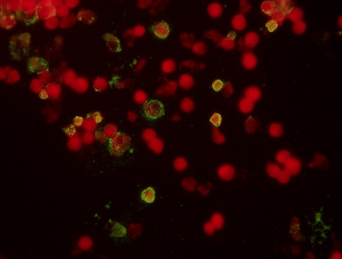
In addition to PCR, the patient's blood can also be serologically tested for the presence of tick-bite fever or spotted fever. However, it often takes more than 5 days until seroconversion. In indirect immunofluorescence, IgM- and IgG antibodies against tick-borne rickettsiae and against spotted fever rickettsiae are determined.

Contact
- Prof. Dr Dennis Tappe
- Research Group Leader
- phone: +49 40 285380-499
- fax: +49 40 285380-252
- email: tappe@bnitm.de
Sample shipment
- Central Laboratory Diagnostics and Medical Care Center
- phone: +49 40 285380-0
- fax: +49 40 285380-252
- email: labordiagnostik@bnitm.de
Examination orders (only in German)
- Examination orders for bacterial disease (only in German) ( PDF 146 KB )


