Dead cells support the functionality and diversity of the immune system's phagocytes
You are what you eat - this also applies to certain phagocytes of the innate immune system, the so-called phagocytic macrophages. In a study, researchers at the University Medical Center Hamburg-Eppendorf (UKE) and at the Bernhard Nocht Institute for Tropical Medicine (BNITM) have discovered that the function and gene expression of macrophages is supported by the dead cells they ingest: The cellular identity of ingested cells contributes to the functional diversity of the phagocytic macrophages. The researchers have published their findings in the journal Science.

The scientists carried out the study using a model of the parasitic infection schistosomiasis, which causes liver damage and is associated with so-called programmed cell death.
"Our results show that targeted feeding of macrophages has the potential to make cell therapies more effective, especially in long-lasting liver diseases such as cirrhosis" says Dr. Lidia Bosurgi, study leader from the UKE’s I. Medical Clinic and Polyclinic and working group leader at the Protozoa Immunology Unit at the BNITM.
The study was funded by the German Research Foundation (DFG) as part of the Collaborative Research Center 841 “Liver Inflammation: Infection, immune regulation and consequences”.
Further information on the Science Paper

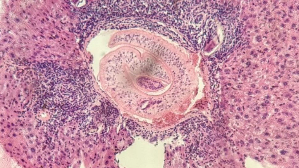
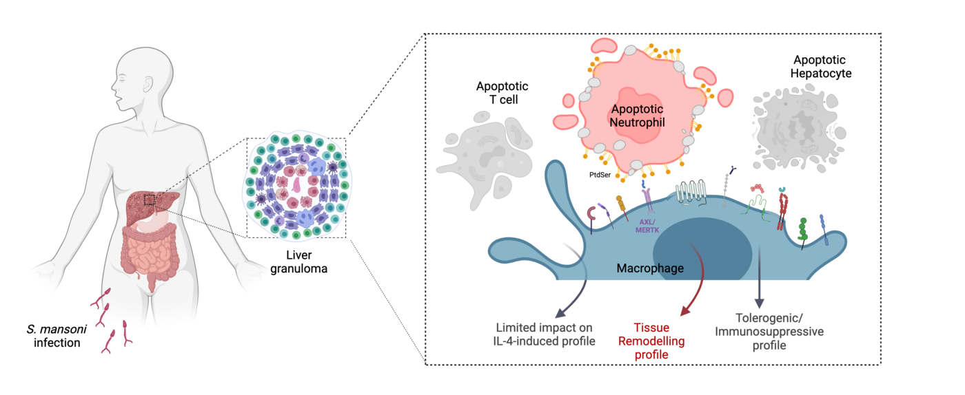
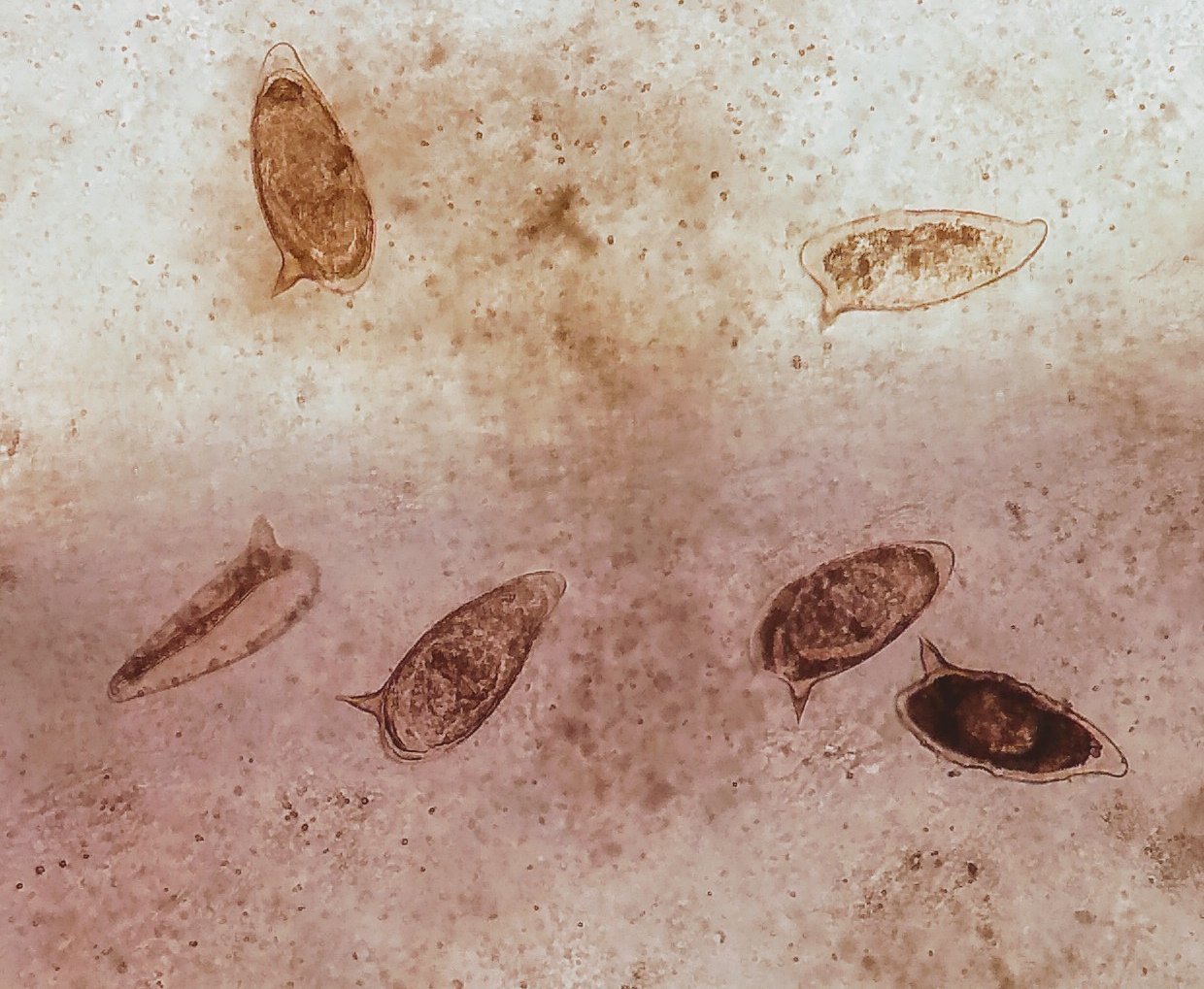
The identity of dying cells phagocytosed influences the function of the phagocytic macrophages. During infection with the parasite Schistosoma mansoni, its eggs become trapped inside the liver of the infected host. Granulomas form around the egg - these are remodeled tissue structures in the liver that contain immune cells such as macrophages in addition to tissue cells. The granuloma environment is enriched with the anti-inflammatory molecule IL-4 (interleukin-4). The granulomas reduce the host's immune response to the egg. The macrophages and various dying cells such as apoptotic T-cells, apoptotic liver cells (hepatocytes) and apoptotic neutrophils (a type of immune cell) come into close proximity in the granuloma.
First author Imke Liebold and her colleagues in Lidia Bosurgi's group carried out the laboratory work at the BNITM. They disscovered that macrophages acquire distinct profiles when ingesting the surrounding dying cells, depending on the cellular identity of the ingested cell: When the macrophages ingest dying neutrophils, they adopt a tissue remodeling profile, which means that they mainly support tissue remodeling. The macrophage receptors AXL and MERTK play an important role in this process - the researchers were able to show that the macrophages did not adopt the tissue remodeling profile when the receptors were experimentally switched off. When macrophages ingest dying liver cells, they subsequently perform tolerogenic/immunosuppressive functions. When macrophages ingest dying T cells, they do not develop a specific profile.

Further information
Link to the original publication in Science: Liebold et al. Apoptotic cell identity induces distinct functional responses to IL-4 in efferocytic macrophages. Science. 2024. DOI: 10.1126/science.abo7027
Contact person
Saskia Lemm
Press officer UKE
Phone : 040 7410-56
Email : presse@uke.de
Dr Anna Hein
Public Relations
Phone : +49 40 285380-269
Email : presse@bnitm.de
Further information


